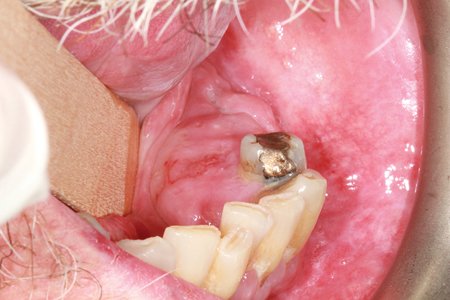
Squamous cell carcinoma is the second most common form of skin cancer in the United States, and it starts in the squamous cells of the epidermis, the outermost layer of skin. More than 1 million people receive this diagnosis each year, and the rate has risen roughly 200% over the past 30 years. Although it forms most often on sun-exposed areas such as the head, arms and legs, it also develops in areas with mucous membranes, including the mouth, lungs and anus.
Recognizing Squamous Cell Carcinoma Symptoms
Squamous cell carcinoma produces noticeable skin changes. A rough bump or growth may crust over like a scab and bleed. Some growths sit higher than the surrounding skin, while a depression forms in the center. Sores also signal the condition. A wound that does not heal, or one that heals and returns, points to a possible problem. A flat, scaly, red patch about 1 inch across may also develop.
Certain skin changes are linked to squamous cell carcinoma. A dry, itchy or scaly bump that differs in color from the surrounding skin is associated with actinic keratosis. A lesion on the lower lip with pale, dry or cracked tissue is associated with cheilitis; it may produce a burning sensation during sun exposure. White or pale patches inside the mouth, on the tongue, gums or cheeks are associated with leukoplakia. Because these changes appear on the surface of the skin or mouth, a healthcare provider should evaluate them.
Understanding Risk Factors
It affects anyone, but certain factors increase the risk. Long-term sun exposure or sun damage early in life raises the likelihood of developing the condition. People with a pale complexion, blue or green eyes, or blonde or red hair also face greater risk. Males develop squamous cell carcinoma at about twice the rate of females; people over age 50 account for the highest number of cases. A weakened immune system, an organ transplant, or exposure to chemicals such as arsenic also increases risk.
Confirming a Diagnosis
A healthcare provider examines the affected area and reviews its size, shape and location. The provider also asks when symptoms began, whether the area changed in size, and whether it causes pain or itching. A skin biopsy removes a small tissue sample for examination under a microscope to confirm cancer cells. Imaging tests, such as a CT scan or MRI, help determine the size of the carcinoma beneath the skin and whether it has spread to nearby lymph nodes. After testing, the provider assigns a stage. Stage 0 remains within the epidermis, while Stage IV indicates the cancer has spread to organs such as the liver, lungs or brain.
Treatment removes cancer from the body, and the approach depends on the size, shape and location of the tumor. Surgical options include excision, curettage and electrodesiccation; surgery is commonly used for cancers on the face. Nonsurgical treatments are also available. Cryosurgery freezes cancer cells, photodynamic therapy combines blue light with light-sensitive agents, and topical creams or immunotherapy drugs treat some cases when surgery is not appropriate.
Schedule Your Skin Exam
Skin changes require medical evaluation. A new lump, a sore that does not heal, or white patches inside the mouth warrant an examination, and a healthcare provider or dermatologist can evaluate these changes. Early diagnosis allows treatment before the cancer spreads beyond the skin because treatment is more effective before the disease advances. Contact a dermatologist to schedule a skin exam.
- Methods for Youthful Skin Rejuvenation
- Functional Health and Its Role in Preventive Care
- Navigating Hormonal Changes and Wellness During Menopause
- Exploring Non-Invasive Treatments for Nerve Pain
- The Role of Dental Exams in Early Detection of Oral Diseases