Abdominal pain can have many causes, and finding the right one often requires a closer look inside. Body imaging tests give your doctor a detailed view of your organs, tissues, and structures. Your provider will explain common imaging techniques used for abdominal pain, while explaining what each one shows.
Create Sound Wave Images
Ultrasound uses sound waves to create images of your abdominal organs, and it produces no radiation. It works well for examining the gallbladder, liver, kidneys, and bladder. Doctors often order it first because it is fast, safe, and widely available. A technician moves a small probe across your skin to perform the body imaging. It detects cysts, masses, and organ enlargement, but dense tissue or gas may limit image clarity. The images appear in real time, allowing your doctor to watch blood flow and organ movement during the scan.
View Abdominal Structures
X-rays are among the fastest imaging tools available. They are often used in emergency settings. They show bones, gas patterns, and potential blockages in the digestive tract. A simple abdominal X-ray takes only minutes, and it yields results almost immediately. While the test is painless, a technician may ask you to stand or lie still while they take the image.
Sometimes your doctor will order more than one view, since different angles can reveal different problems. You usually do not need special preparation, and this makes X-rays convenient and easy to schedule. X-rays use a small amount of radiation, but the dose is generally low. They may be used when doctors suspect a blockage, perforation, or foreign object.
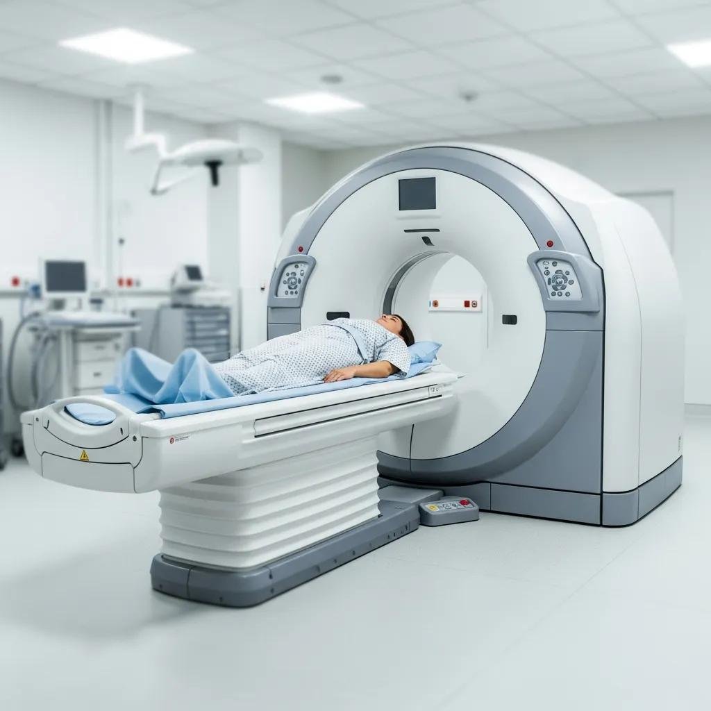
Capture Detailed Cross-Sections
CT scans produce detailed cross-sectional images of the entire abdomen, and they are among the most comprehensive tools available. Body imaging can clearly show organs, blood vessels, lymph nodes, and abnormal growths. Doctors frequently use CT scans when the cause of abdominal pain is unclear or when conditions like appendicitis or a bowel obstruction are suspected. The test is painless, and you will lie still on a table that moves through a ring-shaped scanner. CT scans can also help guide procedures. Before your scan, keep a few key points in mind:
- You may receive a contrast dye to highlight blood vessels and soft tissue more clearly.
- The dye may be given by mouth or through a vein, depending on what your doctor needs to see.
- You may be asked to fast for a few hours beforehand, especially if contrast is used.
CT scans use more radiation than X-rays. This is something to discuss with your doctor. They are fast, and they work well even in urgent situations. If you are pregnant, tell your care team, since they may suggest a different test. You should also mention any allergies before your scan begins. Most people have no discomfort, though the dye can cause a brief warm sensation. The results are often available quickly, and this helps your doctor make timely decisions about your care. CT scans reveal both structural and soft-tissue detail, and they often provide the clearest picture.
Discuss Body Imaging Today
Each imaging technique serves a different purpose, and the right choice depends on your symptoms, medical history, and how quickly a diagnosis is needed. Ultrasound is fast and radiation-free, and this makes it a common first step. X-rays are useful for structural issues, CT scans offer comprehensive detail quickly, and MRI provides the clearest view of soft tissue without radiation. If you are experiencing abdominal pain, speak with your doctor about which imaging test is most appropriate for your situation. Understand your options. Make it easier to move forward.
- FAQs About Hyperbaric Oxygen Therapy for Autism
- The Benefits of Regular Consultations with a Dermatologist
- Debunking Myths About Athlete’s Foot
- What To Know About Diabetes Care
- What To Expect During a Consultation for Colon Cancer Surgery